Dental 2D X-Ray Scans
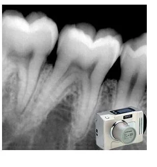
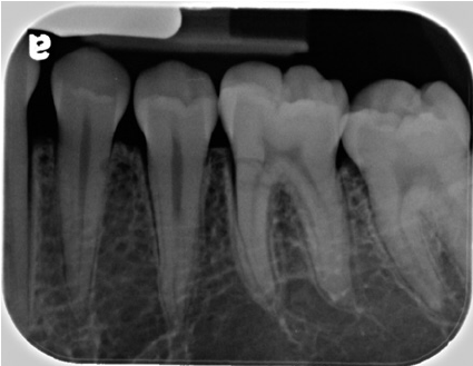
Digital RVG - RadioVisioGraphy
To capture high quality images of teeth & jaw for Real Time Results, Reduced Radiation Exposure, Enhanced Image Clarity, Image Manipulation etc.
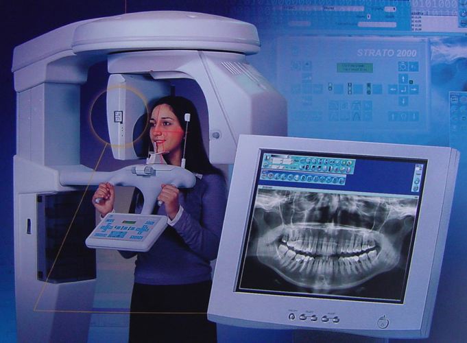
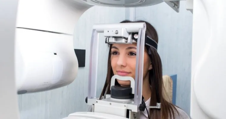
Digital OPG - Orthopantomogram
Shows position, number & growth of all teeth in upper & lower jaw, to diagnose infections, tumors, congenital abnormalities, pre-implant evaluation etc.
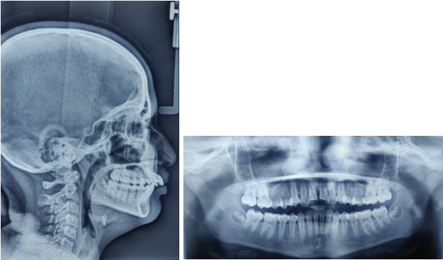
LATERAL CEPHALOGRAM
Shows side of face with very precise positioning so that various measurements can be made to determine current and future relationship of the top and bottom jaw.
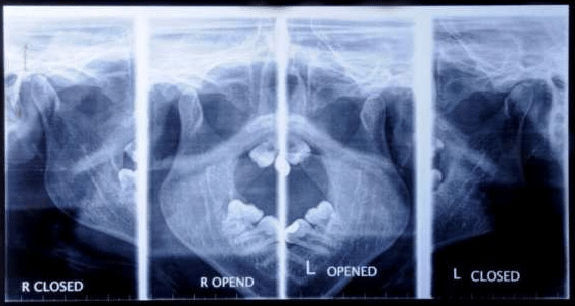
DIGITAL TMJ 4 VIEWS
Identifying structural changes, displaced fractures, assessing excursion and joint spaces in trauma setting, joint noises, trismus and occlusal alterations.
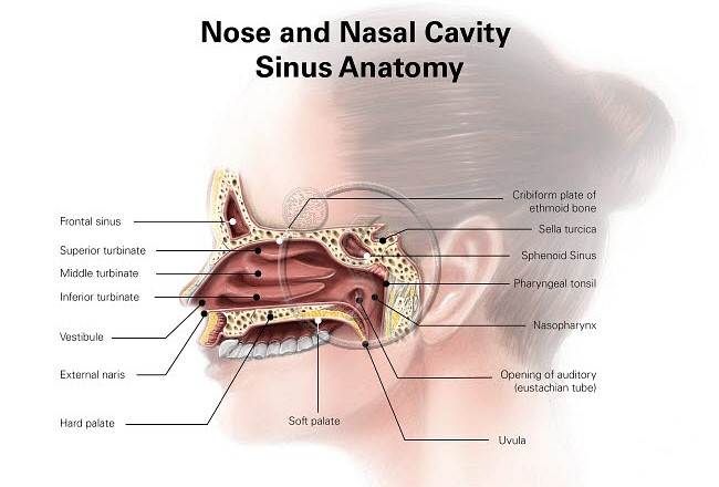
DIGITAL SINUS
Analyzing volumes of left and right maxillary sinuses, nasal & maxillary sinus airway complex, to allow pre-operative assessment of maxillary sinus lift surgery.
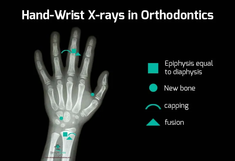
HAND WRIST
To understand the direction of facial development, To predict growth potential left, To clarify a discrepancy between dental age and chronological age etc.

AP VIEW
Anterior-posterior denotes that the central ray passes through the anterior anatomy & exits posteriorly in which posterior structures are closer to the detector.
PA VIEW
Posterior-anterior denotes that the central beam passes from posterior to anterior in which the anterior structures are closer to the detector.
Dental 3D CBCT Scans
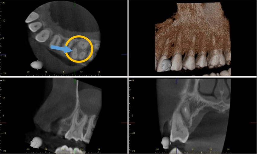
CBCT Single Tooth Scan
CBCT, Cone Beam Computed Tomograph,y creates 3D images that show bone, airway, soft tissue and obtain overall view of entire mouth, jaw, nasal, and throat areas.
CBCT Single Quadrant Scan
Provides detailed information about specific quadrant of the mouth.A C-arm rotates around head, capturing multiple 2D images & combines them to create 3D image.
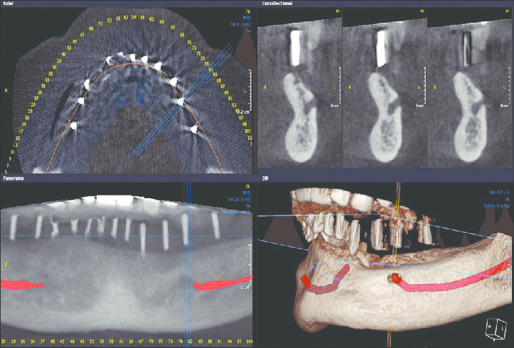
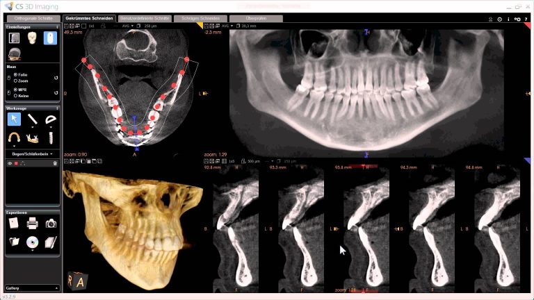
CBCT Mandibular Scan
Provides 3D images of the lower jaw, bone structure, tooth positioning & surrounding anatomy and reduces risk of damaging important structures during surgery.
CBCT Maxillary Scan
Creates detailed 3D images of the upper jaw, teeth & surrounding anatomy. Evaluates the jaw, sinuses, nasal cavity & nerve canal for accurate dental implants.
CBCT Full Mouth Scan
Creates 3D image of mouth, jaw & neck. Shows nasal cavity, tooth decay, tooth root issues etc. Used for planning tooth extractions, root canals etc.
CBCT TMJ Both Scan
Produces 3D images of jaw, nasal & throat areas to diagnose & treat temporomandibular joint (TMJ) disorders by providing detailed images of the TMJ's bone structures.